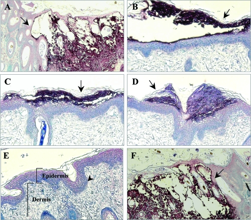
FIG. 7.
Lesion formation in skin xenografts infected with ORF10-to-ORF12 cluster gene mutants. Deparaffinized skin sections harvested at days 21 after infection were incubated with human polyclonal anti-VZV IgG and stained with biotinylated secondary antibody (purple; magnification, ×10). Sections representative of skin xenografts infected with each virus are shown. Large lesions were observed for skin infected with POKA (A) and POKAΔ12 (F). Small lesions (arrows) restricted to the epidermal layer were observed for skin infected with POKAΔ11 (B), POKAΔ10/11 (C), and POKAΔ11/12 (D). POKAΔ10/11/12 (E) did not produce detectable skin lesions. The normal structures of the epidermis, the dermis, and the basement membrane that separates the epidermis from the underlying dermis are indicated in this section.

