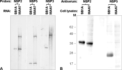
FIG. 2.
Analysis of rotavirus RNAs and expression of proteins in MA/67-infected MA104 cells. Confluent MA104 cell monolayers in a six-well plate infected with either the undiluted MA/67 (0.5 ml/well) or parental SB1A (10 PFU/cell) strain were harvested at 14 h p.i. (A) Viral genomic dsRNAs extracted from the cell lysates were fractionated on a 10% polyacrylamide gel for 10 h at 12 mA and then blotted onto a BrightStar Plus membrane (Ambion Inc., Austin, Tx). The blots were probed with SB1A NSP2-specific (XbaI/EcoRI fragment, 249 bp), NSP3-specific (SpeI/PstI fragment, 597 bp), or NSP5-specific (full-length, 667 bp) cDNA labeled with digoxigenin-11-UTP. Signals were detected by using the DIG High Prime DNA labeling and detection starter kit I (Roche Diagnostics) according to the manufacturer's instructions. (B) Proteins from the harvested cell lysates described for panel A were electrophoresed in NuPAGE gel (Invitrogen) at 200 V for 50 min and then blotted onto nitrocellulose. The blot was probed with guinea pig anti-NSP5 polyclonal antiserum (1:2,000 dilution) or guinea pig anti-NSP2 polyclonal antiserum (1:1,500), followed by treatment with goat anti-guinea pig horseradish peroxidase-conjugated antibody (1:10,000). Molecular mass markers (in kilodaltons) are shown on the left of the gel.

