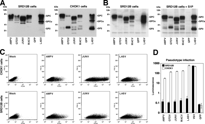
FIG. 3.
S1P is required for processing and function, but not trafficking, of the GPs of human-pathogenic arenaviruses. (A) Expression of arenavirus GPs in S1P-deficient cells. S1P-deficient SRD12B cells and wild-type CHOK1 cells were transfected with flag-tagged GPs of AMPV, GTOV, JUNV, MACV, and LASV or with GFP as a control, and 48 h later, cell lysates were analyzed by Western blotting with an antiflag antibody. (B) Complementation of SRD12B cells with recombinant S1P. SRD12B cells were cotransfected with the indicated flag-tagged GPs and either empty control vector (SRD12B cells) or an expression plasmid for S1P (SRD12B cells + S1P). Viral GPs were detected by Western blotting as described for panel A. (C) S1P processing is not required for cell surface expression of arenaviral GPs. SRD12B cells and CHOK1 cells were transfected with the indicated GPs or, as a control, an empty plasmid (mock). After 48 h, cells were detached by nonenzymatic treatment and live, nonpermeabilized cells were stained with MAb 83.6 to AMPV GP2 and LASV GP2 (21) and MAb BE08 to JUNV GP1 (16). Primary antibody was detected with a phycoerythrin (PE)-labeled secondary antibody and analyzed by flow cytometry using a FACSCalibur flow cytometer (13). Data were acquired and analyzed using Cell Quest and FloJo software packages. In dot plots, the y axis represents forward scatter and the x axis represents PE fluorescence intensity. (D) S1P-mediated processing is required for the function of arenavirus GPs. SRD12B cells and CHOK1 cells were cotransfected with a plasmid expressing MLV gag and pol, the MLV genomic plasmid pLZRS-Luc-gfp, and expression plasmids for the indicated GPs, and 48 h later, conditioned supernatants were harvested, cleared, and added to Vero E6 monolayers. Infection was detected by a luciferase assay as described in the legend for Fig. 2D. Note that the y axis has a log scale.

