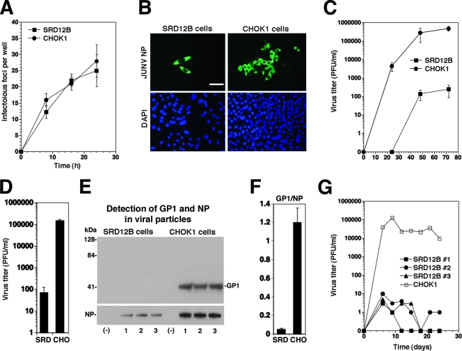
FIG. 5.
S1P processing is required for the production of infectious JUNV and cell-to-cell propagation. (A) Infection of SRD12B and CHOK1 cells with JUNV Candid 1. Cells (96-well plates) were infected with 100 PFU of JUNV Candid 1. At the indicated times, cells were fixed and stained with MAb BG12 for JUNV NP. The number of infectious foci was determined in each well (n = 3; error bars indicate standard deviations [SD]). (B) Representative infectious foci in SDR12B and CHOK1 cells at 48 h postinfection. Bar = 20 μm. (C) Production of infectious JUNV in SRD12B and CHOK1 cells. Cells were infected with JUNV Candid 1 at an MOI of 1. Cell supernatants were harvested at the indicated times, and infectious virus titers were determined by a plaque assay on Vero E6 cells. A compilation of three independent experiments is shown (means ± SD). (D to F) Analysis of the composition of JUNV particles produced in SRD12B and CHOK1 cells. Triplicate samples of 5 × 10⁶ SRD12B (SRD) and CHOK1 (CHO) cells grown in T75 flasks were infected with JUNV Candid 1 at an MOI of 1. (D) Cell culture supernatants were harvested after 48 h, and infectious virus titers were determined by a plaque assay on Vero E6 cells. (E) Detection of viral proteins by Western blotting. Supernatants were subjected to ultracentrifugation through a 20% sucrose cushion (6). Pellets were solubilized in hot SDS-PAGE loading buffer, and viral proteins were separated by SDS-PAGE under nonreducing conditions. Blots were probed with MAbs GB03-BE08 (anti-JUNV GP1) and SA02-BG12 (anti-JUNV NP) (16) combined with a biotinylated secondary antibody and horseradish peroxidase-conjugated streptavidin. Signals were detected using the Super Signal West Femto chemiluminescence detection kit from Pierce. (F) Densitometric analysis of the blots in panel E. Densitometry was performed as described in the legend to Fig. 4C, and the ratios of the signal intensities for GP1 (IGP1) to NP (INP) were calculated (GP1/NP1) for virion particles from SRD12B cells (SRD) and CHOK1 cells (CHO). (G) Persistent infection of SRD12B cells with JUNV Candid 1 does not result in escape variants. SRD12B (three independent cell cultures) and CHOK1 cells were infected with JUNV Candid 1 at an MOI of 1 and passaged every 3 days. At the indicated times, virus titers were determined by a plaque assay. The results of one representative example out of three independent experiments is shown.

