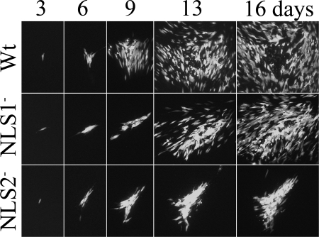
FIG. 3.
NLS2− mutant spreads more slowly than either WT virus or the NLS1− mutant. HFF cells were infected with low multiplicities of WT virus or the NLS1− or NLS2− mutant. All viral genomes contained an ectopic copy of the GFP gene as a marker for the presence and replication of viral DNA. Shown are fluorographic images of the cultures taken 3, 6, 9, 13, and 16 days after infection. The same plaques were photographed on each of the five days. Note comparatively larger WT focus on days 6 to 16, loss of fluorescent cells from the center of WT focus by day 16, and comparatively high density of fluorescent cells in the NLS2− focus on days 9 to 16.

