FIG. 6.
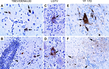
Localization of viral antigens in the CNS of monkeys inoculated with TBEV/DEN4Δ30 at 7 dpi (A and B), with LGTV at 14 dpi (C and D), or with YF 17D at 7 dpi (E and F). (A) Frontal cortex, showing strongly immunolabeled neurons (arrows) within intact gray matter. (B) Thalamus, showing immunolabeled neurons (arrows) surrounding a perivascular inflammatory infiltrate (arrowhead). (C and D) Ventral horns of the lumbar spinal cord, showing neuronophagia (D) and prominent immunolabeling in the cytoplasm of degenerating neurons (C and D, arrows). (E) Parietal cortex, showing neuronophagia (arrow) and strongly immunolabeled neurons with decorated processes. (F) Putamen, showing CII (arrowhead) and prominent immunolabeling of neuronal network (arrows). Original magnifications, ×400 (A to E) and ×200 (F).

