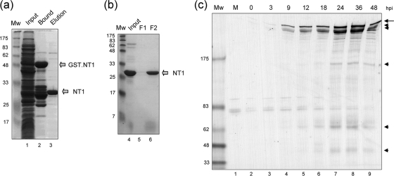
FIG. 1.
VP1-2 NT1 fragment purification and antibody generation. (a) Total protein staining of the soluble fraction of bacterial extracts expressing the GST-NT1 fusion protein (lane 1), the bound, purified GST-NT1 fusion protein (lane 2), or the final NT1 protein after specific protease cleavage and elution (lane 3). This last sample was then loaded onto a Mono Q column and fractionated as described in Materials and Methods. The Mono Q input and sample fractions are shown in panel b. Peak fractions were pooled and used to immunize rabbits. (c) Vero cells were mock infected or infected with the HSV-1 17 strain. At different times postinfection, samples were analyzed by Western blotting with the antibody raised against NT1, αVP1-2NT1r. The long arrow indicates full-length VP1-2, with cleavage products indicated by arrowheads.

