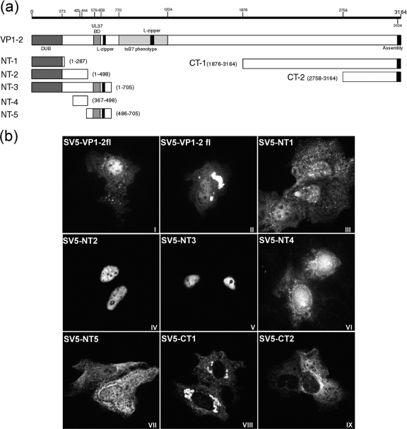
FIG. 3.
Cellular localization by immunofluorescence of transfected VP1-2 and VP1-2 fragments. (a) Schematic illustration of HSV-1 17 strain VP1-2 indicating positions of certain published features (including information on the PrV homologue), from the N-terminal end (NT) to the C-terminal end (CT): the deubiquitination enzymatic domain (DUB) (1 to 273), the putative NLS (425 to 444) region from the present work, the UL37 binding domain (UL37 BD) (579 to 609), putative leucine zipper sequences (L-zipper), a SalI fragment mapped as the tsB7 phenotype (770 to 1204), and the CT end involved in nuclear assembly (3104 to 3164). Subregions expressed from the various constructs are illustrated, including amino acid boundaries. (b) Monolayers of Vero cells were transfected with the plasmids showed in panel a. The cells were fixed with methanol 24 h after transfection and analyzed for localization using the monoclonal antibody against the SV5 N-terminal epitope tag. Representative images are shown for each fragment, including two examples of VP1-2 full length (VP1-2fl).

