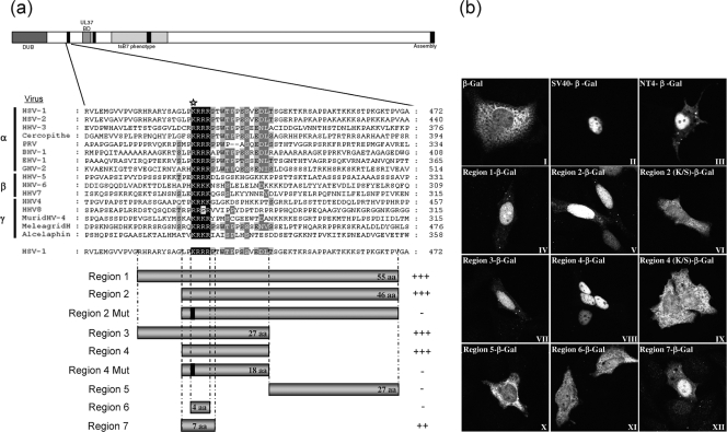
FIG. 4.
A functional NLS in VP1-2 transferable to a heterologous protein. (a) Schematic diagram illustrating the alignment of the N-terminal motif of VP1-2 from the three different herpesvirus families. Regions encompassing the highly conserved basic region of HSV VP1-2 were fused to the β-Gal gene, as illustrated in the bar diagrams below. Region 2 and region 4 were also analyzed, with a single substitution in a conserved lysine (K→S), represented as a solid line within the corresponding schematic and by a star on the sequence line. (b) Monolayers of Vero cells were transfected with the plasmids expressing the different regions of VP1-2 NT4 as β-Gal fusion proteins. Cells were fixed with methanol 24 h later and analyzed for β-Gal localization. Representative images are shown for each fusion region. Summary conclusions on the efficiency of the candidate regions are illustrated next to the schematic in a semiquantitative manner: +++, very efficient nuclear localization (at least as efficient as that of the SV40 NLS-β-gal vector); ++, mainly nuclear localization but also some weaker cytoplasmic distribution of the signal; -, no significant nuclear accumulation.

