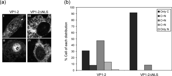
FIG. 5.
Effect of NLS deletion on localization of VP1-2. (a) COS cells were transfected with 1 μg of the pcDNA3-SV5-UL36fl (panels I and II) or pcDNA3-SV5-UL36ΔNLS (panels III and IV) expression vector. After 24 h, the coverslips were fixed and VP1-2 localization examined using the monoclonal antibody against the V5 tag. Panel b shows the distribution of the percentages of cells with localization patterns of VP1-2 or VP1-2ΔNLS, defined as follows: only cytoplasmic (C) or only nuclear (N); equal distribution in both compartments, C = N; cytoplasmic more abundant than nuclear, C>N; nuclear more abundant than cytoplasmic, C<N. At least 100 cells were counted. VP1-2 distribution was mixed, with a significant percentage of cells showing cytoplasmic and nuclear distribution or nuclear distribution greater than cytoplasmic. The effect of the NLS deletion was clear, with the majority of cells exhibiting cytoplasm-restricted localization.

