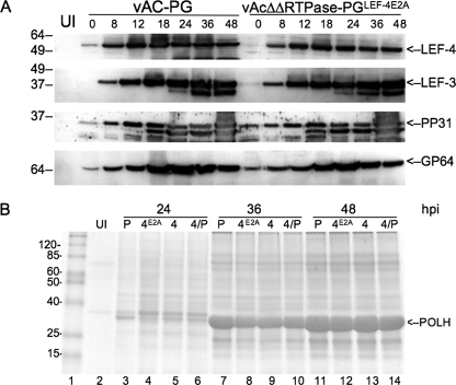
FIG. 4.
Western blot analysis of viral protein expression. (A) Sf-9 cells were infected with the double-mutant virus vAcΔΔRTPase-PGLEF-4E2A or the control vAc-PG at an MOI of 5. Cells were harvested at the indicated times, and detergent-soluble protein extracts were separated on SDS gels. Each lane represents 1 × 10⁴ cells. The lane labeled UI represents uninfected cells. Time zero is the end of a 1-h adsorption period. Extracts were probed with antibody against rabbit antiserum raised against LEF-4 or PP31 or mouse monoclonal antibodies against LEF-3 or GP64. (B) Analysis of polyhedrin expression in cells infected with vAC-PG (P), vAcΔΔRTPase-PGLEF-4E2A (4A), vAcΔΔRTPase-PGLEF-4 (4), or vAcΔΔ RTPase-PGLEF-4/PTP (4/P). Cells were harvested at the indicated times and detergent-insoluble extracts were separated on 12% acrylamide gels and stained with Coomassie brilliant blue.

