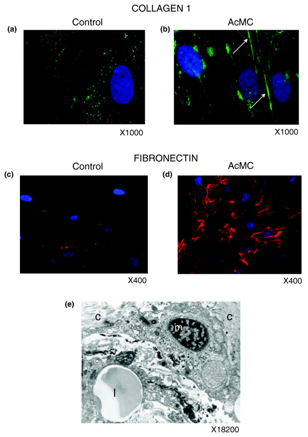
Figure 14.
Macrophages promote ECM component secretion by pre-adipocytes. (a-d) Immunofluorescence analysis of pre-adipocytes cultivated with control (a,c) or AcMC-conditioned media (b,d) reveals that they express type 1 collagen (b, green staining, arrows) and fibronectin (d, red staining) in the latter condition. (e) Electron microscopy picture of an adipose tissue macrophage ('m') shows its collocation with collagen fibers ('c') and a lipid droplet ('l'). (a-d) Representative images of five independent experiments.

