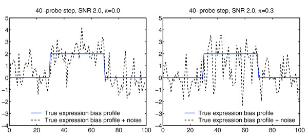
Figure 1.
Simulation model. Blue solid: Original gene expression bias profile containing a centrally located region with increased expression. Black dotted: Corresponding gene scores, generated by mixing a high-frequency signal component into the original bias profile (details in Materials and methods). Left: Example signal generated with 40-probe step with SNR 2.0, and no non-influenced genes (π = 0.0). Right: Corresponding signal with a higher proportion of non-influenced genes (π = 0.3).

