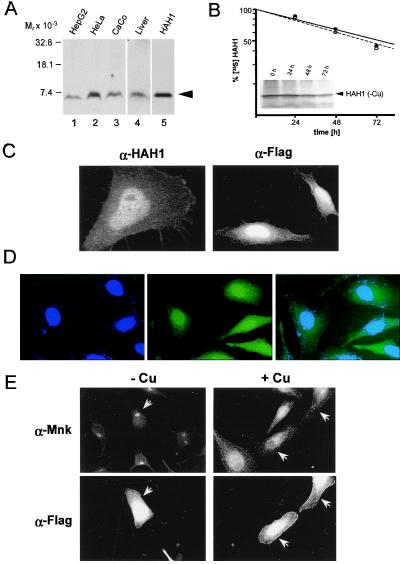
Figure 1.
(A) Immunoblot analysis with HAH1 antibody. Protein lysates (100 μg) (lanes 1–4) or 100 ng of recombinant HAH1 expressed in E. coli (lane 5) was separated by SDS/PAGE on 10–20% Tricine gradient gels, transferred to nitrocellulose, and analyzed by chemiluminescence. (B) HeLa cells grown in the presence of BCS (●) or copper (○) were lysed and immunoprecipitated with HAH1 antisera. Following a 2-hr pulse with [35S]methionine and [35S]cysteine, cells were lysed and immunoprecipitated at indicated chase times followed by SDS/PAGE, autoradiography, and quantitation of bands. (Inset) HAH1 immunoprecipitated in the presence of BCS. (C) Immunofluorescent localization of HAH1 in HeLa cells incubated with HAH1 antisera (α-HAH1; confocal 100X) and Flag M2 antibody (α-Flag; 60X). (D) Immunofluorescence of HeLa cells stained for nucleus with DAPI (narrow-band UV), HAH1 antiserum (FITC), or overlay images of DAPI and HAH1. (E) Double immunofluorescence in transfected HeLa cells treated with BCS (−Cu) or copper (+Cu) followed by incubation with Menkes (α-Mnk) and Flag-M2 (α-Flag) antibodies. Arrows indicate transfected HeLa cells.

