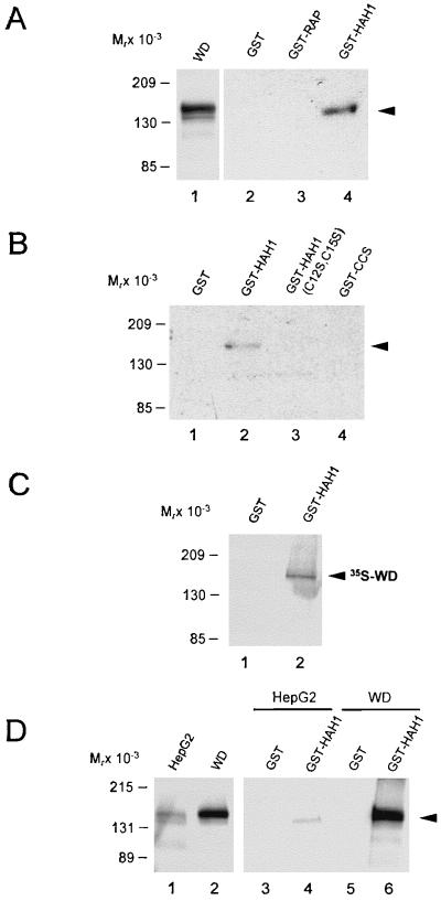
Figure 2.
In vitro interaction of HAH1 and Wilson protein. (A) COS-7 cell lysates (150 μg) expressing Wilson protein (5 μg; lane 1) were incubated with 50 pmol of purified GST-fusion proteins (lanes 2–4), and eluates were subjected to SDS/PAGE, transferred to nitrocellulose, and examined by immunoblotting with Wilson antisera. (B) COS-7 cell lysates expressing the Wilson protein were incubated with the indicated GST constructs (lanes 1–4) and analyzed as indicated above. (C) Full length Wilson cDNA was translated in vitro in the presence of [35S]methionine and [35S]cysteine followed by incubation with the GST constructs as indicated. Interacting proteins were separated by SDS/PAGE and analyzed by autoradiography. (D) One milligram of HepG2 cell lysate (100 μg; lane 1) or 150 μg COS-7 cell lysate expressing the Wilson protein (5 μg; lane 2) was incubated with 3 nmol of purified GST fusion proteins (lanes 3–6) and analyzed by immunoblotting with Wilson antisera as before.

