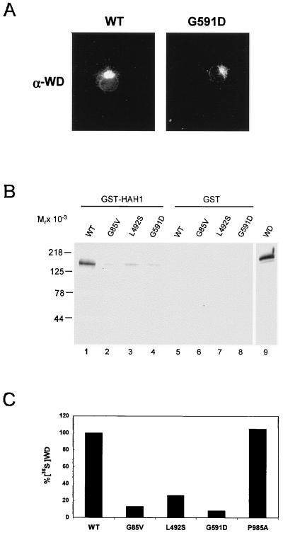
Figure 5.
(A) Indirect immunofluorescence of Wilson protein in COS-7 cells transiently transfected with wild type (Left) or G591D mutant (Right). Cells were processed for immunofluorescence 72 hr posttransfection (X60). (B) COS-7 cells expressing either wild-type or mutant Wilson proteins were lysed, incubated with GST-HAH1, and interacting proteins were separated by SDS/PAGE and analyzed by immunoblotting with Wilson antisera. (C) Wild-type and mutant Wilson proteins were translated in vitro in the presence of [35S]methionine and [35S]cysteine, incubated with GST-HAH1, and interacting proteins analyzed by fluorography after SDS/PAGE and were quantitated by PhosphorImager.

