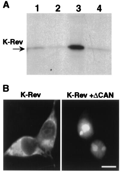
Figure 1.
Functional interaction between Crm1 and K-Rev. (A) Microaffinity columns containing GST (lane 1), GST-Crm1 only (lane 2), GST-Crm1 preincubated with Ran-GTP (lane 3), or GST-Crm1 preincubated with Ran-GDP (lane 4) were prepared as described (6, 28). Equal aliquots of 35S-labeled K-Rev were then loaded onto each column, and bound proteins were eluted by using 0.05% SDS. The eluates were resolved by gel electrophoresis and visualized by fluorography. (B) Human 293T cells (35-mm cultures) were transfected with 500 ng of pBC12/CMV/K-Rev and 1 μg of pBC12/CMV/ΔCAN or the parental pBC12/CMV plasmid by using the calcium phosphate procedure. At ≈48 h after transfection, the cells were fixed and stained as described (6) by using a 1:500 dilution of a rabbit polyclonal anti-K-Rev antiserum and a 1:2,000 dilution of a FITC-conjugated donkey anti-rabbit antiserum (The Jackson Laboratory). No staining was observed in mock-transfected cultures. (Bar ≈20 μm.)

