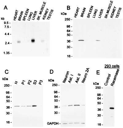
Figure 3.
Distribution of serine racemase. (A) Northern blot analysis. A full-length serine racemase probe was used to probe a multiple-tissue rat mRNA membrane (CLONTECH). (B) Western blot analysis of serine racemase in rat tissues. Immune serum to serine racemase was used at 1:10,000 dilution. (C) Subcellular fractionation of brain extracts showing enrichment of serine racemase in the cytosolic fraction. H, total homogenate; P1, 9,000 × g pellet; P2, 30,000 × g pellet obtained from P1 supernatant; S3, supernatant of 120,000 × g pellet representing soluble cytosolic proteins; P3, 120,000 × g pellet. (D) Enrichment of serine racemase in glia. Western blot of serine racemase in neuronal culture virtually free of glia, astrocyte type 1 primary culture (Ast. I), astrocyte type 2-enriched culture (Ast. II), and neuroblastoma (Neuro 2A cells). To assure that the same amount of protein was loaded per lane (20 mg homogenate protein), the blot was reprobed for glyceraldehyde-3-phosphate dehydrogenase (GAPDH). (E) Western blot of serine racemase in transfected HEK293 cells. Each lane contains 100 ng of homogenate protein.

