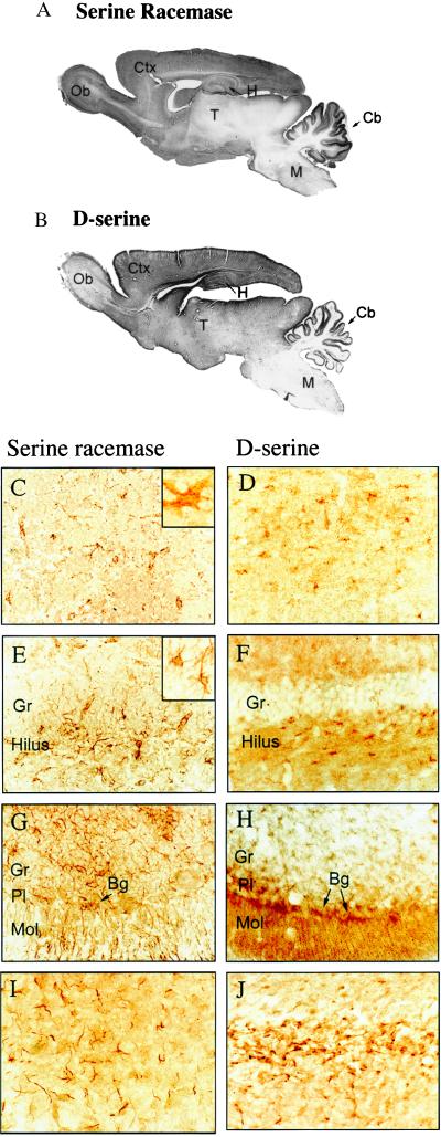
Figure 4.
Colocalization of serine racemase and endogenous d-serine in brain. (A) Immunohistochemical staining for serine racemase. (B) Immunohistochemical staining for d-serine. In the cerebral cortex (C and D), both serine racemase and d-serine are evident in glial cells with morphology corresponding to astrocytes. (C, Inset) A ×1,000 magnification of a glial cell positive for serine racemase in the cerebral cortex. In the hippocampus (E and F), several glial cells containing serine racemase and d-serine occur in the hilus of the dentate gyrus. Granule cell neurons (Gr) are unstained. (E, Inset) A ×650 magnification of glial cells positive for serine racemase in the hippocampus. In the cerebellum (G and H), Bergmann glia cells (Bg) in the Purkinje cell layer (Pl) and scattered stellate astrocytes and glial processes in the granule cell layer (Gr) are positive for both serine racemase and d-serine. Arrows depict Bergmann glia cell bodies that extend processes toward the pia in the molecular layer (Ml) of the cerebellum. In the corpus callosum (I and J), astrocytes are strongly labeled for both serine racemase and d-serine. Though both serine racemase and d-serine occur in the same types of cells, subtle morphologic differences are noticeable, perhaps reflecting the use of different fixatives and tissue processing for each staining. Except for the insets, magnifications are ×400.

