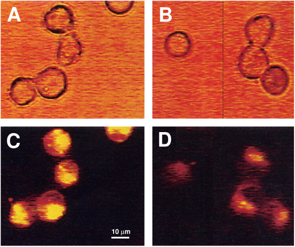
Figure 2.
Fluorescent and light microscopic analysis of alteration in intracellular K+ concentrations in H9 cells treated with recombinant Nef. H9 cells were loaded with the fluorescent indicator PBFI-AM and incubated with 300 nM recombinant Nef protein or with control medium without Nef for 15 min at 37°C. Control cells (A and C) and cells incubated with Nef protein (B and D) were examined by light (A and B) and fluorescent (C and D) microscopy.

