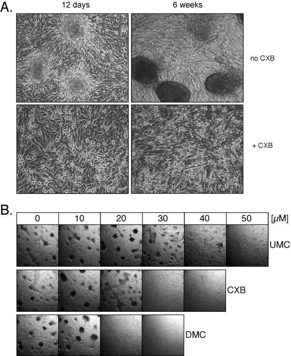
Figure 4.
Prevention of focus formation by CXB, DMC, and UMC. U251 glioblastoma cells were continuously exposed to various concentrations of CXB, DMC, or UMC for up to 3 months in the same cell culture dishes (i.e., without splitting the cell monolayers). In A., cells were treated with or without 30 μM CXB and photomicrographs (160× magnification) were taken after 12 days and 6 weeks. Top left shows early-stage focus formation (3 representative foci are shown); top right shows 3 examples of fully developed, very compact foci (each one consisting of an estimated several hundred cells). Neither early-nor late-stage foci were present in CXB-treated cell cultures. In B., cells were treated with increasing concentrations of the three drugs and photomicrographs (20× magnification) were taken after 10 weeks. Note that in the absence of drug treatment, many foci (several hundred per square inch) had developed. However, no foci developed in the presence of 20 or 30 μM DMC, 30 or 40 μM CXB, and 50 μM UMC (representative sections of each culture are shown).

