
Figure 3.


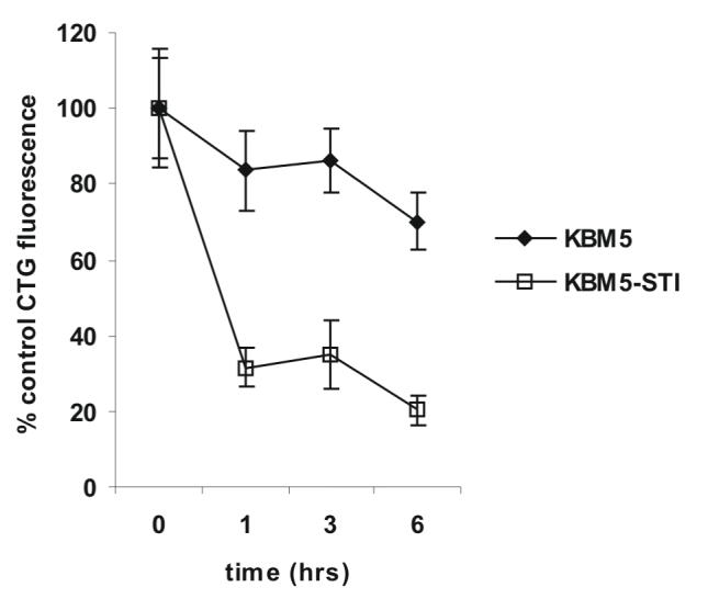
CDDO-Me induces rapid generation of ROS, mitochondrial dysfunction, and oxidation of intracellular GSH. A) Cells were treated with CDDO-Me as indicated and the generation of intracellular ROS was quantitated by flow cytometry as described in the Materials and Methods. B) Cells were loaded with the potentiometric probe TMRM, treated with CDDO-Me as indicated, and ΔΨM was quantitated by flow cytometry as described in the Materials and Methods. C) Cells were treated with CDDO-Me as indicated and loaded with the GSH specific probe Cell Tracker Green ™ followed by incubation in ice for 10 min. Cells were then extensively washed in PBS, and the GSH specific fluorescence of CTG was measured as described in the Materials and Methods.