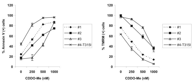
Figure 5.


CDDO-Me induces GSH depletion and apoptosis in primary CML blast crisis cells. A) Cells obtained from patients in blast crisis CML were cultured ex vivo with increasing concentrations of CDDO-Me (0-1000 nM) for 24 h and viable cells were counted after trypan blue staining using a hemocytometer. B) Patient samples were treated with increasing concentrations of CDDO-Me (0-1000 nM) for 3 h and intracellular GSH was quantitated by flow cytometry as described in the Materials and Methods. C) Patient samples were treated as in A) and phosphatidyl serine externalization and ΔΨM were quantitated by flow cytometry as described in the Materials and Methods.

