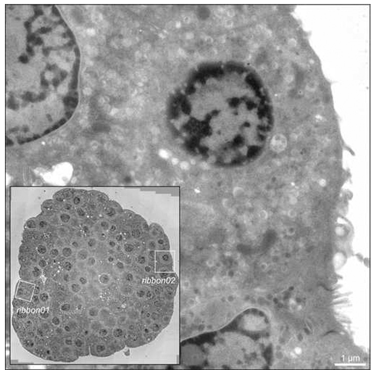
Fig. 1.
Intact islets of Langerhans isolated from adult mice were cultured overnight in vitro, stimulated with elevated extracellular glucose (11 mM) for 60 mins, and preserved for EM by high-pressure freezing, freeze-substitution and plastic embedding. Inset: 2D survey showing the whole islet in cross-section, acquired by stage-shifted montaging of 12×12 images at 4700× magnification using the data acquisition program SerialEM (Mastronarde, 2003; Mastronarde, 2005). Boxed regions highlight the two islet beta cells (designated ‘ribbon01’ and ‘ribbon02’) in the islet imaged and reconstructed by ET. Main image: 0° (untilted) view taken from one of the tilt series of islet beta cell ribbon02. Scale bar: 1 µm.

