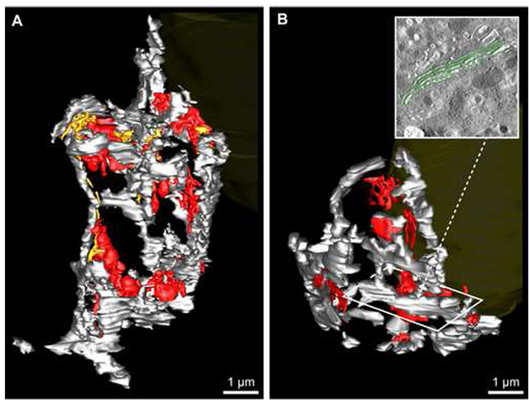
Fig. 5.
The 3D organization of the entire Golgi ribbon was analysed in both glucose-stimulated islet beta cells reconstructed in toto. Membranes of the Golgi complex were segmented in ribbon01 (A) and ribbon02 (B) to distinguish between the main Golgi ribbon (i.e. the ‘ribbon proper’), comprised of stacked cis- and medial cisternae (grey), the trans-most Golgi cisterna (red) and the penultimate trans-cisterna (gold). The nucleus (yellow) is partially visible in the figure background for reference; the orientation of the Golgi relative to the cell is the same for ribbon01 and ribbon02 as for Fig. 3 and Fig. 4, respectively. B, inset: A portion of the segmented stacked cis-medial Golgi cisternae (highlighted in green) shows how the main Golgi ribbon was segmented.

