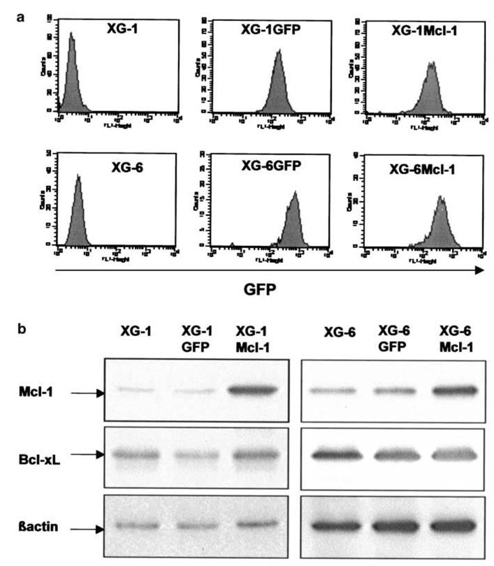
Figure 4. Transduction of myeloma cells with Mcl-1-GFP retrovirus or control GFP retrovirus.

A: XG-1 and XG-6 myeloma cells were transduced with Mcl-1-GFP or GFP retroviruses and were selected with 700 μg/mL G418 and 2 ng/mL IL-6. All selected myeloma cells highly expressed GFP after 15 days of culture.
B: Parental XG cells, XG cells transduced with Mcl-1-GFP or GFP retroviruses were cultured with IL-6 and harvested for Western blot analysis. Blots were probed with anti-Mcl-1, anti-Bcl-xL or anti-β actin antibodies. Western blots are for one representative experiment out of three.
