Figure 7.
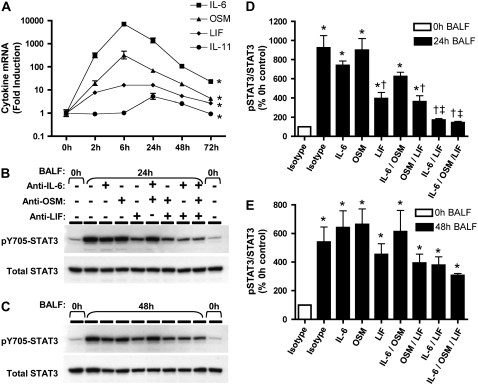
IL-6 family cytokines and pneumonia. IL-6 family mRNA in the lungs (A) was measured using real-time RT-PCR for IL-6, oncostatin M (OSM), leukemia inhibitory factor (LIF), and IL-11 mRNA. Pneumonic lung lobes were harvested from C57BL/6 mice 0 to 72 hours after intratracheal E. coli, and fold-induction values (versus uninfected mice) are expressed as geometric means ± geometric SE (n = 5–6). *Statistically significant effect of infection on mRNA levels for all shown (P < 0.05). IL-6 family-dependent STAT3 activation in MLE-15 cells (B–E) was measured using immunoblots. MLE-15 cells were stimulated for 10 minutes with BALF collected 0, 24 (B/D), or 48 hours (C/E) after intratracheal E. coli, in the absence and presence of neutralizing antibodies for IL-6, OSM, and/or LIF. Representative images from one of three separate experiments are shown for pSTAT3 and total STAT3 immunoreactivity in MLE-15 cell protein extracts in response to pooled BALF collected at (A) 24 and (B) 48 hours. Immunoreactive bands shown were approximately 90 kD in size. The two rows in panels B and C show the same membrane labeled first for pSTAT3 and the second, after stripping, for total STAT3. pSTAT3 immunoreactivity was quantified by densitometry (D–E) and normalized to that of total STAT3. pSTAT3/STAT3 ratios are expressed as a percentage of the value determined for MLE-15 cells stimulated with control BALF (0 h BALF) and expressed as means ± SE (n = 3). *Statistically significant compared with cells stimulated with 0 hours of BALF and isotype control IgG (P < 0.05). †Statistically significant compared with cells stimulated with pneumonic BALF and isotype control IgG. ‡Statistically significant compared with cells stimulated with pneumonic BALF and anti-LIF (P < 0.05).

