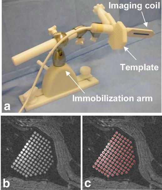
FIG. 1.

Needle placement and imaging device. a: The needle-guiding template is fixed at a right angle to the endorectal imaging coil. After positioning, both are fixed in place with an immobilization arm. b: The template holes, filled with surgical lubricant, are easily visualized in MR images. c: After registration of the position and orientation of the needle-guiding template, colored dots (representing the path of each needle hole) are projected through the image volume. Visualization of the template allows for easy verification of this registration.
