
FIG. 3.
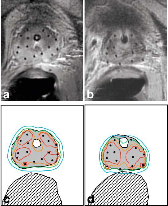
High-dose-rate (HDR) brachytherapy catheter placement and isodose maps. At the beginning (a) and end (b) of a 5-week course of external beam radiation therapy, HDR brachytherapy was performed using catheters placed under MRI guidance (both images are from the same patient). c,d: Radiation isodose maps, corresponding to a,b, indicate 150% (red contour), 125% (orange contour), 100% (green contour), and 75% (blue contour) of the prescribed radiation dose (1050 cGy). The prostate (gray filled region), urethra (white region inside the prostate), and rectum (hatched region) are also shown. Note that the green, 100% dose contour conforms well to the prostate margin, while overdose of the urethra and rectum is avoided.