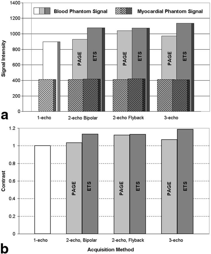
FIG. 5.
a: Comparison of signal intensities for blood (back, solid) and myocardial phantoms (front, hatched). All methods were able to increase blood phantom signal intensities while the myocardial signal remained constant. b: Comparison of contrast, normalized to that of single-echo SSFP. Contrast increased slightly for all implementations of multishot EPI-SSFP, compared to single-echo SSFP. ETS produced slightly higher blood and myocardial SIs than PAGE, leading to slightly better contrast values as expected from the use of higher flip angles allowed by longer TRs.

