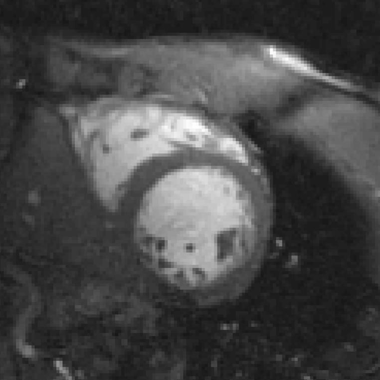
FIG. 7.
High-resolution (256 × 192, rectangular FOV) end-diastolic image acquired with two-echo bipolar SSFP, PAGE, and 180 T · m-1 · s-1 slew rates. Higher resolution allows for clearer visualization of the papillary muscle while maintaining excellent blood-myocardium contrast. Note that the susceptibility artifact in the posterior-lateral wall is more prominent due to extended TR.

