
Fig. 7.
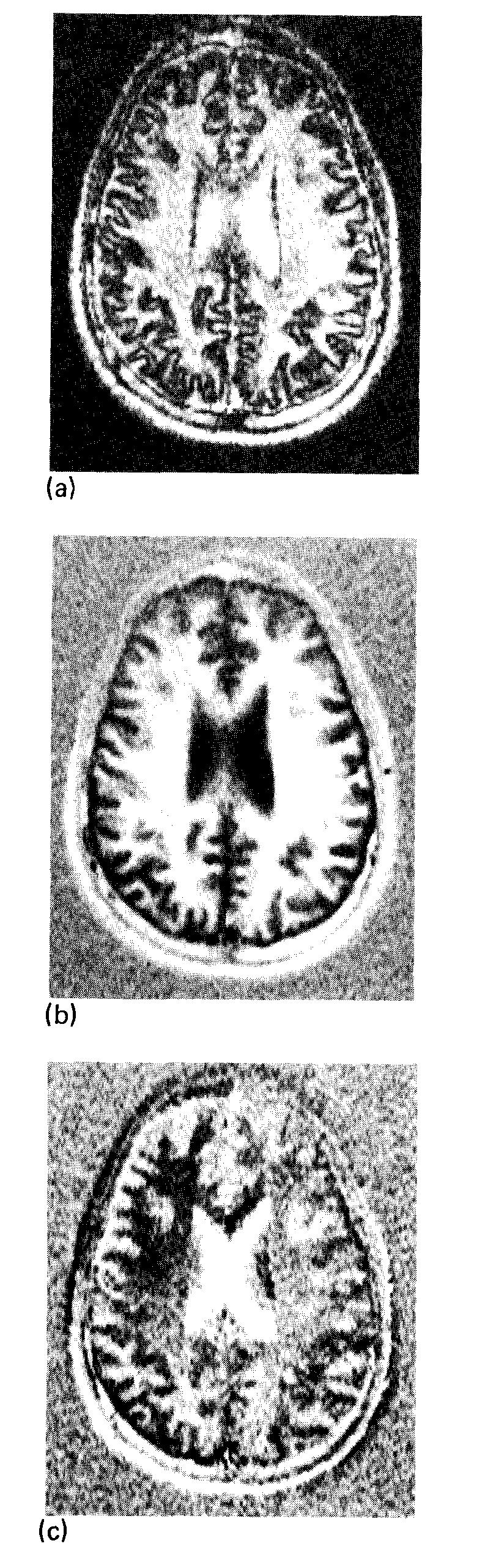
Transverse images of a 1-cm slice through the head obtained with an ISE sequence (TR = 2000 ms, TI = 240 ms, TE = 30 ms). (a) Using magnitude reconstruction, phase information is lost and regions of long TI, such as CSF and GM, have inverted, positive signal intensity. (b) The “real” component of the complex image. This image is phase sensitive. (c) The “imaginary” image is the projection of the image vectors onto the axis orthogonal to that in (b). The window for (b) is 20 times the background root-mean-square deviation, the window for (a) and (c) is 10 times the background rms deviation of (b).