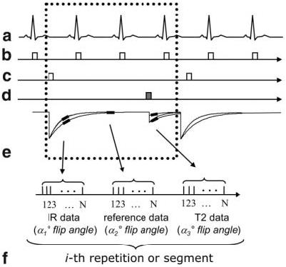
Figure 2.

Pulse sequence diagram for MCODE with acquisition of T1-weighted IR data, reference data for phase-sensitive image reconstruction, and T2-weighted data: (a) EKG, (b) R-wave trigger, (c) 180° inversion pulses, (d) T2-preparation, (e) magnetization, and (f) data acquisition (during mid-diastole) for the i-th repetition or segment.
