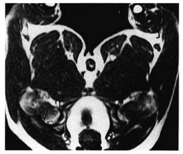
Fig. 2.

An axial T1-weighted spin-echo image at the level of the femoral head and neck in a normal dog (TR/TE = 500/20). The orientation is the same as in Figure 1. Marrow is predominantly fatty in the proximal femoral head (small open arrow) and predominantly hematopoietic distal to the physeal line (large open arrow).
