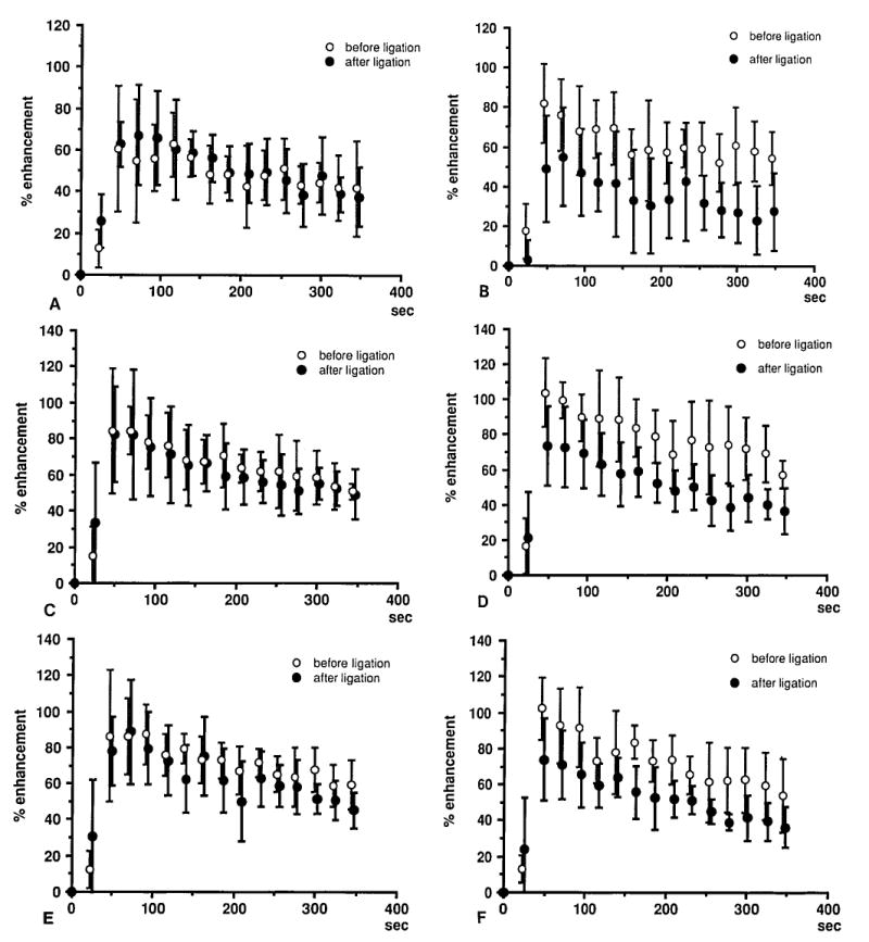
Figs. 4A-4F.

Percent enhancement versus time after Gd injection for each anatomic region. Curves before and after unilateral ligation are compared. A rapid rise followed by a slower decrease is seen. There is 25% to 45% reduction in percent enhancement after venous occlusion on the ligated side. Proximal portion of the femoral head on the right (control) (A) side and left (ligation) side. (B) Distal portion of the femoral head on the right (control) (C) side and left (ligation) (D) side. Femoral neck on the right (control) (E) side and left (ligation) (F) side.
