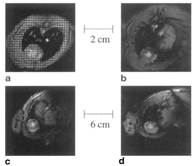
FIG. 4.

MRIs of the heart acquired with four coils. (a) Helmholtz rat coil, (b) cosine rat coil, (c) Helmholtz rabbit coil, (d) cosine rabbit coil. Only the rat Helmholtz image is tagged. The infarction is located on the lower left side of image (a). The other three images are taken from sham animals and hence do not show any infarct.
