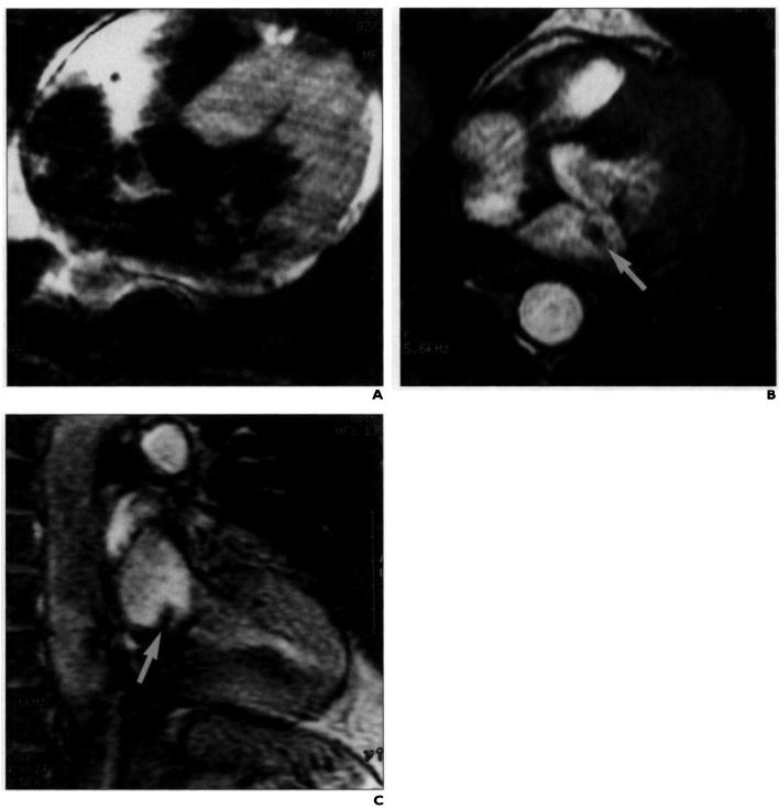
Fig. 2.
Cardiac valve assessment using segmented K-space acquisition in 67-year-old man with supravalvular mass on echocardiogram. Patient was referred for MR imaging for characterization of relationship between left atrial mass and mitral valve. A-C, Spin-echo T1-weighted MR image (A) and segmented K-space acquisitions in axial (B) and sagittal (C) planes reveal fixed thrombus in left atrium (arrow, B and C). Acquisition time for A was 5 min 43 sec. Each cine sequence, one frame of which is shown in B and C, was acquired in 14 heartbeats and consisted of 15 cine frames using eight views per segment.

