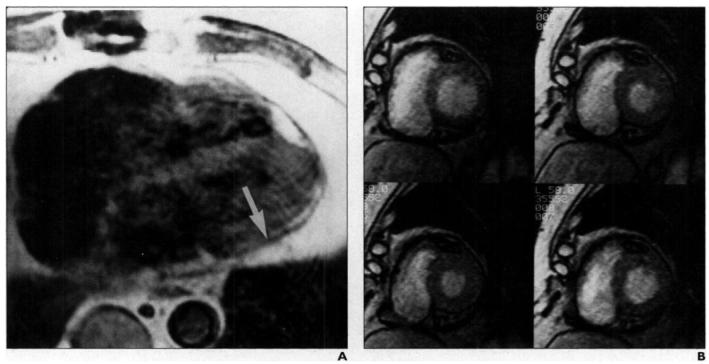
Fig. 4.
Cardiac wall motion evaluation using segmented K-space acquisition in 59-year-old man evaluated for possible constrictive pericarditis after cardiac transplant.
A, T1-weighted spin-echo image shows normal thickness of pericardium (arrow).
B, Short-axis images of left and right ventricles show cine frames at 45, 175, 305, and 435 msec after R wave (displayed left to right, top to bottom) obtained in 16 heartbeats with eight views per segment. Normal function and diastolic filling of both ventricles were noted on cine review. A myocardial biopsy subsequently revealed evidence of rejection.

