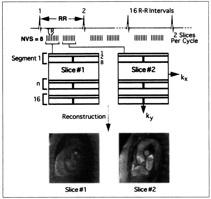
Fig. 6.
Diagram summarizes segmented K-space gradient-recalled echo acquisition in a multislice, single-phase mode. Each R-R interval is again divided into short intervals, each yielding number of views per segment, except each interval now corresponds to different slice location, each obtained during different portion of cardiac cycle. In this example, one slice location is obtained during systole, and another is obtained during diastole. This technique provides for rapid vascular screening of entire aorta in one breath-hold. Because flow-related enhancement is greater during systole, using only first portion of cardiac cycle increases vascular signal, at expense of slightly increased image acquisition time. NVS = number of views per segment.

