FIG. 4.

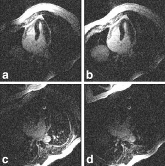
Magnitude images for individual coils illustrating B1 weighting of ghost artifacts. The artifact from CSF in the spinal cord is evident in images from the back coils (c and d), and is suppressed in chest coil images (a and b).

Magnitude images for individual coils illustrating B1 weighting of ghost artifacts. The artifact from CSF in the spinal cord is evident in images from the back coils (c and d), and is suppressed in chest coil images (a and b).