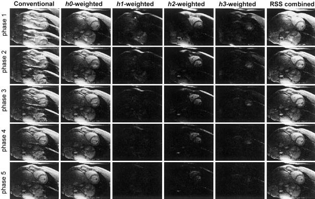
FIG. 5.
Images comparing conventional image reconstruction with ghosts (left column) and PAGE reconstructed images (right columns) for ECG triggering segmented SSFP acquisition during initial cardiac phases (no magnetization preparation). The individual separated ghost image components are shown in columns 2-5. The ghost-cancelled RSS combined magnitude images (excluding h3-weighted images of column 5) are shown in right the column.

