
FIG. 3.
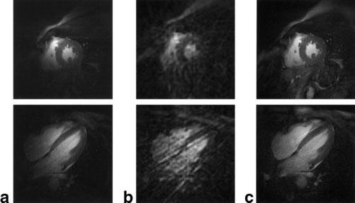
End-systolic images in mid short-axis (top row) and four-chamber long-axis (bottom row) orientations. The images in column a were reconstructed using only the signal acquired with the small loop coil. The low-resolution images (12 views) in column b are representative of those used for ROI correlation mask h(x,y) selection, and the images in column c are the corresponding full-resolution images reconstructed using 144 total views and the signals from five coils.