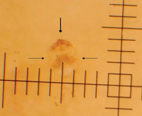
Figure 4: Photographic image of a female scabies mite taken through a hand-held epiluminescent stereomicroscope (original magnification × 40). Note the hang-glider-like triangle of the mite's head (vertical arrow) and round body (horizontal arrows). The hash marks on the axes are at 0.1-mm intervals.
