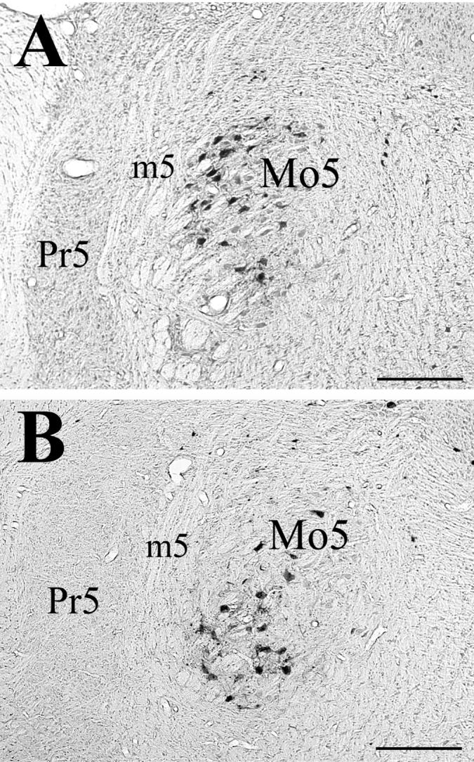
Figure 1.

Photomicrographs of labeled motoneurons in the (A) rostral and (B) caudal portions of the trigeminal motor nucleus, from an animal that survived 72 hrs following PRV-BA injections into ipsilateral masseter muscle. Abbreviations: m5, motor root of 5th nerve; Mo5, trigeminal motor nucleus; Pr5, principal sensory nucleus. Scale bars represent 500 μm.
