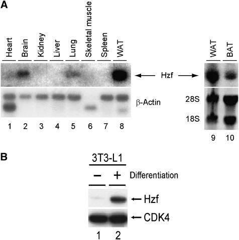
Figure 1.
Hzf is highly expressed in adipose tissue. (A) Expression of Hzf mRNA was analysed in normal mouse tissues using Hzf cDNA as a probe. WAT (white adipose tissue) and BAT (brown adipose tissue) were obtained from epididymal and interscapular adipose, respectively. (B) Expression of Hzf protein in 3T3-L1 cells (−: undifferentiated, +: differentiated for 2 days) was analysed by immunoblotting. CDK4 was used as a loading control.

