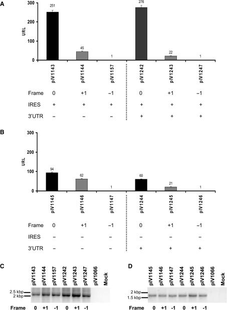
Figure 2.
Translation of chimeric HCV core-luciferase sequences in frames 0, +1 and −1 in hepatoma cells. Plasmids containing nts 342–822 of the core coding sequence fused to a luciferase gene reporter cDNA were transfected into Huh-7 cells. The translation of HCV 5′ sequences was studied in the three frames [0 (core frame) in black, +1 (ARFP frame) in gray and −1 frame in white] and in the presence (A) or absence (B) of the IRES sequence, using luciferase activity quantification. The integrity of the RNA transcripts was investigated in Huh-7 cells transfected with HCV-luc plasmids containing the IRES (C) or lacking the IRES (D). RT-PCR of HCV-luc transcripts. Total RNA was purified from Huh-7 cells transfected with the indicated plasmids and reverse transcribed as described earlier. The resulting cDNAs were amplified by PCR using HCV (Forward 1 and 2) and Luc-specific primers (Reverse 1, see Figure 1A for primer position). Plasmid pIV1066 encoding luciferase and Huh-7 cells transfected with an irrelevant plasmid were used as controls.

