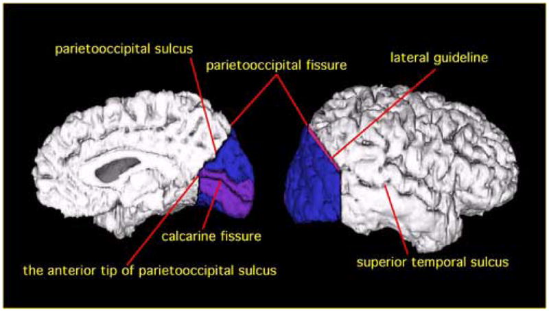
Figure 1.

Delineation of PVA and VAA in occipital lobe. PVA is shown in purple, and VAA is shown in blue. For the medial surface (left), the border between parietal lobe and occipital lobe is the parietooccipital sulcus. PVA is defined as the area including one gyrus above and one gyrus below the calcarine fissure. For the lateral surface (right), the border between the two lobes is delineated by a guideline connecting the parietooccipital fissure and the superior temporal sulcus on the most anterior slice of occipital lobe (The guideline is shown in red).
