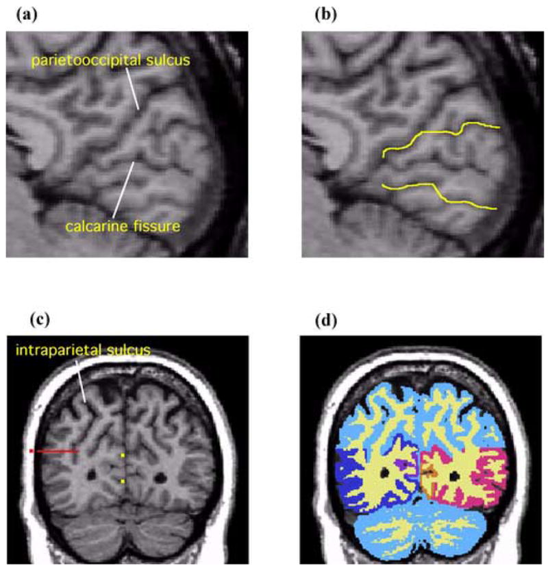
Figure 2.

Sagittal and coronal MR images showing delineation of PVA and VAA. (a) The rater identifies the parietooccipital sulcus and the calcarine fissure on the midsaggital plane. (b) The yellow lines are the guidelines extending the sulcal courses used to delineate PVA and VAA. (c) On a coronal slice PVA and VAA are delineated by referring to the guidelines (yellow dots). In part B, here shown as yellow dots. On the lateral surface, the parietal lobe and occipital lobe are operationally separated by extending the guideline (the red dot) horizontally and medially across the tissue bridge of white matter horizontally and medially up to the intraparietal sulcus. (d) A coronal view of PVA and VAA delineation. White matter and gray matter were shown in light yellow and light blue respectively. The gray matter of PVA is shown in orange (subject left) and purple (subject right). The gray matter of VAA is shown in red (subject left) and blue (subject right).
