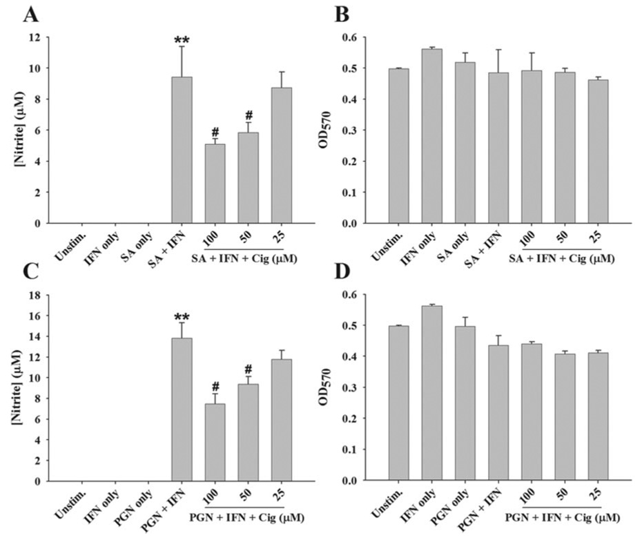
FIGURE 11.
Ciglitazone interferes with the priming effects of IFN-γ in S. aureus-stimulated microglia. Primary microglia were seeded in 96-well plates at 2 × 10⁵ cells per well and incubated overnight. The following day, cells were first pretreated with the indicated doses of ciglitazone (final concentrations) for 1 h followed by 100 ng/ml recombinant mouse IFN-γ for 1 h. Subsequently, microglia were stimulated with heat-inactivated S. aureus (107 CFU/well) (A and B) or PGN (10 µg/ml) (C and D). At 24 h following bacterial exposure, cell-free supernatants were collected and analyzed for NO expression (A and C). Microglial viability was assessed using a standard MTT assay and the raw OD570 readings are reported (B and D). Results are reported as mean ± SD of three independent wells for each experimental treatment. **, p < 0.001 significant differences between microglia exposed to S. aureus or PGN only compared with cells treated with bacterial stimuli plus IFN-γ #, p < 0.05 significant differences between microglia exposed to S. aureus or PGN plus IFN-γ vs cells pretreated with the various concentrations of ciglitazone plus S. aureus or PGN plus IFN-γ. Results are representative of three independent experiments.

