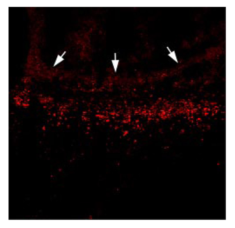
FIGURE 7.
PPAR-γ immunoreactivity is localized along the developing brain abscess wall. Serial 10-µm thick sections were prepared throughout brain abscesses collected at day 6 post-infection, subjected to immunofluorescence staining for PPAR-γ expression (red), and imaged by confocal microscopy at a magnification of ×10. PPAR-γ immunoreactivity was detected within the peri-abscess tissue, where the developing abscess wall is demarcated (arrows). Results are representative of two independent experiments using a total of n = 10–12 individual mice.

